AOU/COS/SCO 2008 Meeting in Portland: Part I

This week was the joint conference of the American Ornithologist's Union, the Cooper Ornithological Society and the Society of Canadian Ornithologists in Portland, Oregon. This meeting celebrated the 125th anniversary of the American Ornithologist's Union(AOU) and ushered in a new president for the AOU, Dr. Edward "Jed" Burt who is a faculty just down the road from Cincinnati in Delaware, OH at Ohio Wesleyan University. It was a great meeting and many important ties were forged between Cincinnati Museum Center and researchers and natural history museums around North America and the world. I meet with friends and colleagues from the University of Windsor, University of Alaska at Fairbanks, the Taiwan Endemic Species Research Institute, University of Cincinnati, Auburn University, the Delaware Museum of Natural History, the Cleveland Museum of Natural History, and many other colleges, universities and museums and promoted greater use of the collection and plotted out new collaborations and research projects.

There were many excellent talks on the latest findings in ornithology and my next blog will provide a survey of some of the highlights, but, perhaps most useful was a nearly day long symposium on avian museum collections that included live demonstrations of the latest preparatory methods from some of the top curators and collections managers in the country. This symposium in particular proved to be invaluable and provided me with a wealth of information from preparation to permits that will greatly improve the collection at Cincinnati Museum Center. Also, meeting with colleagues and forging new ties resulted in several new projects. The plan is have in house research at Cincinnati Museum Center result in at least 10 new publications over the next year. A bold goal but one that can be achieved through the numerous collaborative efforts between Cincinnati Museum Center Zoology Department and top researchers in avian biology from around the globe.

Of course I take every opportunity to increase the collection at Cincinnati Museum Center and collected many digital photos to go into a growing georeferenced digital resources database for birds. The meeting reinfornced the utility of this growing type of natural history collection in several talks regarding the ORNIS distributed database system. I gained new insight during this part of the meeting on how to manage these collection and provide proper georeferencing (location data critical to making a useful digital resources collection). Also I learned of new ways in which digital resources in ornithological collections are being used alongside both traditional material (skins, skeletons, spread wings, etc.) and frozen tissue collections. Shown in this blog entry are three new digital photos to be archived in a growing digital resources database for ornithology (Top, Northern Fulmar; middle, Barred Owl; bottom, Pacific-slope Flycatcher).

-END

On the flip side...

ResearchBlogging.org

Critics of evolution often point to a perceived lack of transitional forms in the fossil record as evidence that some forms of life don't share a common genetic heritage but rather arose independently. Creationists have been using this line of argument for over a hundred years. But, they do so in spite of the evidence. Intermediate forms are those organisms that have a mosaic of characteristics linking two seemingly unrelated groups or an organism that exhibits a character state in between those found in two or more other organisms. Creationists claim these forms are lacking in the fossil record. However, the transition between theropod dinosaurs and modern birds, the evolution of the mammalian ear bones from a reptilian jaw, the evolution of whales from terrestrial mammals and even the fossil record for our own species are all classic examples of evolutionary transitions complete with several intermediate forms.

Add to the growing list of evolutionary transitions another example from the fossil record. Vertebrate animals have a bilateral body plan, that is they exhibit a front and back and dividing the body down the middle results in two symmetrical sides. There are few exceptions to the symmetrical vertebrate body plan. One striking exception familiar to us all are the flatfishes (Order: Pleuronectiformes). This group includes fish found at your local market or seafood restaurant including sole and halibut. Adult flatfishes are asymmetrical. They start off life with the typical symmetrical body found in other fishes however as they grow their skull undergoes a radical developmental change with the eyes rotating to one side of their head. A flatfish on the sea bed is therefore a fish essentially lying on it's side with an asymmetrical head. Imagine lying on your left side with both your eyes on the right half of your face and you'll get the picture.

The question is how did this unusual body plan evolve? There are clear benefits to exploiting an open ecological niche and becoming specialized to be a bottom feeder but doing so can mean a radical change in an organism's body plan. The flatfish body plan clearly evolved from a typical symmetrical fish plan. Comparing the details of flatfish anatomy and their genes shows that they fit within the large radiation of bony fishes, nearly all of which have a symmetrical body plan. Also, the turbots (Psettodes sp.) are the living representatives of the earliest branch of the flatfish family tree and it, as expected if flatfish evolved from a symmetrical ancestor, they have one eye that doesn't quite make it all the way around the head during development. What's more, the adult asymmetrical flatfish plan develops from a symmetrical larval body plan. Together all these data indicate that the asymmetrical flatfish plan evolved from a symmetrical ancestral body plan. However, as creationists are fond to ask, where are the intermediates?

Along comes University of Chicago evolutionary biologist Matt Friedman. In the July 10th edition of the journal Nature Friedman provides evidence for the evolutionary transition between symmetrical bony fishes and the asymmetrical flatfishes. Friedman provides highly detailed descriptions of fossil fishes in the genus Amphistium and describes a new species, Heteronectes chaneti. These fossils were available to researchers before, however, Friedman applied computed tomography to the specimens to obtain detailed three-dimensional images of their anatomy. Computed tomography involves moving an x-ray source and detector around a specimen and digitally reconstructing a detailed three-dimensional image from the x-ray exposures. Previously it was difficult to tell an asymmetrical flatfish-style skull from a symmetrical skull crushed by the weight of sediment during fossilization. With the new imaging technology Freidman concluded that these fossil fish do indeed show different intermediate stages leading to the fully asymmetrical modern flatfish body plan. Specifically, the part of the skull containing the orbits, the neurocrania, rotates over evolutionary time. A close relative of the flatfishes, Trachinotus, shows the classic symmetrical condition with one eye on either side of the head. The fossil Amphistium and Heteronectes show an intermediate stage with one eye squarely on one side of the head and the other eye on the other side of the head but shifted up towards the top of the head. The earliest branch on the flatfish family tree, the turbots, one eye rests nearly on top of the head and in the rest of the flatfish the eye has migrated fully to the other side of the head.

New findings are popping up all the time from fields ranging from paleontology to developmental genetics that affirm the conclusion that life shares a common ancestry and that these unusual body plans, such as the asymmetrical head of a flatfish, have arisen through evolutionary processes.

Friedman, M. (2008). The evolutionary origin of flatfish asymmetry. Nature, 454(7201), 209-212. DOI: 10.1038/nature07108

-END

Top ten new species for 2007

ResearchBlogging.orgIt is astonishing just how many critters are out there who are to date unknown to science. Even well known groups like birds and mammals occasionally have a new species described. But for large groups, like insects, marine invertebrates, or plants, finding new species is much more common, particularly in little studied areas like tropical forests or deep sea marine habitats. The International Institute for Species Exploration at Arizona State University has released it's list of the top ten new species for 2007 (click here for photos of these amazing critters). These include a new species of fruit bat from the Philippines (Esselstyn 2007), a mushroom discovered on the campus of Imperial College in London (Taylor et al. 2007), a bright pink millipede from Thailand (Enghoff et al. 2007) and an electric ray from South Africa whose genus name is reminiscent of a popular brand of vacuum cleaners (Compagno and Heemstra 2007). Discovering new species is just one of the many potential exciting aspects of museum-based zoology!

ESSELSTYN, J.A. (2007). A New species of stripe-faced fruit bat (Chiroptera: Pteropodidae: Styloctenium) from the Philippines. Journal of Mammalogy, 88(4), 951. DOI: 10.1644/06-MAMM-A-294R.1

TAYLOR, A., HILLS, A., SIMONINI, G., MUNOZ, J., EBERHARDT, U. (2007). Xerocomus silwoodensis sp. nov., a new species within the European X. subtomentosus complex. Mycological Research, 111(4), 403-408. DOI: 10.1016/j.mycres.2007.01.014

ENGHOFF, H.,SUTCHARIT, C.,PANHA, S. (2007) The shocking pink dragon millipede, Desmoxytes purpurosea, a colorful new species from Thailand (Diplododa: Polydesmida: Paradoxosomatidae). Zootaxa, 1563, 31-36.

COMPAGNO, L. J. V.,HEEMSTRA, P. C. (2007) Electrolux addisoni, a new genus and species of electric ray from the east coast of South Africa (Rajiformes: Torpedinoidei: Narkidae), with a review or torpedinoid taxonomy. Smithiana Bulletin, 7, 15-49.

-END

Butterflies, nanotech and BugFest 2008

ResearchBlogging.org

This weekend was the Cincinnati Museum Center's annual BugFest. Together with my son Cameron we had a great time showing visitors a sampling of butterflies from the zoology department's entomology collection (see photo left). Public presentations of museum collections should introduce the role of museum collections in scientific research. Museum collections and other collections-based field work can have uses that will come as a surprise to many. Focusing on butterflies Cameron and I chose to present the role collections can play not only in gaining a basic understanding of nature but also their potential role in very practical applications in materials science.

High magnification images of the wing of a Morpho butterfly were fed to an LCD screen and used to show visitors the intricate scales that make up a butterfly wing. The very fine micro-structure of these wing scales is what creates the iridescent blues and greens of butterfly wings. While many colors in nature are due to pigments embedded within biological structures like hair, feathers or scales other colors are purely structural created by the particular scatter of light reflected from a structured compound like keratin in birds' feathers or layers of chitin in insect scales. Many blues and greens in bird feathers and insect scales tend to be determined by structure rather than pigments. The very fine microscopic structure of a blue feather or scale therefore determines the wavelengths of light it reflects and thus it's color.

Believe it or not a knowledge of how nature produces colors is useful in nanotechnology. Nanotechnology deals with the engineering of very tiny machines on the size scale of a cell. Mimicking nature can be very useful in producing components for these tiny nano-devices. Butterfly wing scales are studied by engineers to create nano-parts with very particular optical properties. Jingyun Huang et al. in 2006 in the journal Nano Letters found that the wing scales of the iridescent blue butterfly Morpho peleides could be used as a template for making tiny artifical scales of aluminum oxide. These artificial aluminum oxide butterfly scales had identical reflective properties to their natural counterparts and they could be used in nanotechnology to split beams of light. Radislav Potyrailo et al. in the journal Nature Photonics in 2007 published a paper describing the ability of the wing scales of Morpho sulkowskyi to act as components in tiny optical gas sensors.

Of course museum collections act as accessible storehouses of biological diversity and the holdings in collections, like those at Cincinnati Museum Center, can provide material for numerous applications (many of which would have never been anticipated by the original collectors) in both basic and applied research.

Potyrailo, R.A., Ghiradella, H., Vertiatchikh, A., Dovidenko, K., Cournoyer, J.R., Olson, E. (2007). Morpho butterfly wing scales demonstrate highly selective vapour response. Nature Photonics, 1(2), 123-128. DOI: 10.1038/nphoton.2007.2

Huang, J., Wang, X., Wang, Z. (2006). . Nano Letters, 6(10), 2325-2331. DOI: 10.1021/nl061851t

-END

Can you pick up a drop of water with tweezers?

ResearchBlogging.org
Some birds can! A group of shorebirds called phalaropes have a curious way of feeding. They feed on the surface of lakes, tidal wetlands and other bodies of water by swimming around in a tight circle on the surface furiously kicking their legs. This creates a vortex in which tiny aquatic invertebrates like shrimp, aquatic insects and copepods are pulled to the surface. Once food items are trapped in the swirling water phalaropes gobble them up with their long thin bills. But, while the phalarope's curious means of concentrating aquatic invertebrates is well known less well understood is how they use their bills to consume their tiny water suspended prey. A team at the Massachusetts Institute of Technology lead by Manu Prakash has uncovered just how suspension feeding birds like phalaropes use their bills to draw up droplets of water packed with their invertebrate prey.

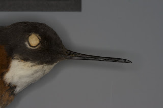
Unlike a straw a bird's bill is open on both sides and opens in an up-and-down motion much like a pair of tweezers so they can't suck up shrimp filled pond water like you would a slushie. In phalaropes their bills are long and very thin (see photo to the right of a Red-necked Phalarope (Phalaropus lobatus) from the Cincinnati Museum Center's Zoology collection). Just how phalaropes can use a tweezer-like bill in a straw-like fashion is a puzzle. Prakash and colleagues found that a drop of water in a very thin bill can be drawn up the bill by what they call a "capillary ratchet". They looked at data in the literature derived from real bills (much of which was originally from museum specimens) and built a mechanical bill with similar properties. When the bill is closed the drop of water is compressed and when opened again it moves a bit further up the bill towards the mouth. Close the bill again the water is compressed. Open again and it moves a bit further up the bill. Click HERE for a Quicktime movie of the Prakash et al. mechanical bill in action. The ability of an artificial bill to serve as a capillary ratchet is highly dependent on both it's shape and it's wetting properties.

This study has several important implications. First, it describes a novel evolutionary adaptation in birds and helps us better understand the myriad of solutions that evolutionary processes can generate to basic challenges in life. Second, an understanding of biomechanics for natural structures like the bill of the phalarope can help human engineers design devices for moving very small amounts of liquid (i.e. microfluidic transport systems). Such devices, inspired by nature, can have important implications in nanotechnology and molecular biology and could potentially advance human health. Finally, because Prakash et al. found that the wetting properties of the bill were critical in its ability to act as a capillary ratchet device environmental managers should look for effects of pollutants on the feeding efficiency of phalaropes and other shorebird species. Petroleum products and detergents could have significant effects on the wetting properties of a phalarope bill and in turn lead to less food for affected birds.

Of course museums, like Cincinnati Museum Center, often play a significant role in these biomechanical studies by serving as storehouses of all the clever tricks invented by evolutionary processes. Taping into nature's diversity is not only good simply for the sake of a greater knowledge of our living world but it also can provide us with designs for our own technology, designs that have been tested over eons of evolutionary tinkering.

Prakash, M., Quere, D., Bush, J.W. (2008). Surface Tension Transport of Prey by Feeding Shorebirds: The Capillary Ratchet. Science, 320(5878), 931-934. DOI: 10.1126/science.1156023

-END

The weird and wonderful platypus genome

ResearchBlogging.org

Modern comparative biology has truly entered a new age. The list of species for which researchers have completely sequenced their genomes continues to rapidly grow. Fruit flies (Drosophila melanogaster), chickens (Gallus gallus), sea urchin (Strongylocentrotus purpuratus), pufferfish (Fugu rubripes), Short-tailed Opossum (Monodelphis domestica), mosquitos (Anopheles gambiae), Rhesus Macaque (Macaca mulatta), several plants such as rice (Oryza sativa) and cottonwood (Populus trichocarpa), numerous microbes, and even Humans (Homo sapiens) all have complete genome sequences completed.

Add to that list the Duck-billed Platypus (Ornithorhynchus anatinus). A research team has just completed the first sequencing of the platypus genome. The platypus is truly among the strangest of mammals. Found exclusively in Australia and Tasmania, they have hair and produce milk as do the rest of their mammalian kin but they also lay eggs and have a brain much like a reptile. Male platypus also sport a spur on their hind feet that can deliver a venomous sting. Because of this odd mix of reptilian and mammalian characters the first platypus specimens brought back by the early explorers of the Australian continent were thought to be a hoax, patched together from bits and pieces of other animals. Cincinnati Museum Center's Zoology Collection has an old platypus specimen in it's holdings (see photo left).

Like it's reproductive behavior, physiology and morphology the genome of the platypus reveals it's key evolutionary position at the base of the mammal family tree. For example, mammalian ova have an outer membrane called the zona pellucida which aids in fertilization. Of the proteins make up the zona pellucida in mammals four found in the platypus match those found in the human genome, however, the platypus genome has two additional ova membrane proteins previously found only in birds. Additionally, the platypus genome contains genes for the yolk protein vitellogenin, a protein found in the eggs of birds but neither marsupials or placental mammals.

The genes underlying the venom found in the spurs of male platypus also tell an interesting evolutionary story. Platypus venom, like many venoms found in reptiles, is a complex mix of different proteins. Platypus venom contains 19 different compounds. The venom proteins in platypus venoms appear to have arisen through duplications of genes. Gene duplication is a common evolutionary process that can give rise to new characteristics. When a gene is duplicated the new duplicate is free to accumulate new mutations and take on new functions while the original gene retains it's original function. Not only has gene duplication played a role in the evolution of platypus venom but the same process likely led to the evolution of venoms in reptiles. Also, the venom proteins in the platypus arose from the same gene families as in venomous reptiles providing an interesting case of convergent evolution (evolution of similar traits arising independently in different lineages).

The complete sequence of the platypus genome follows previous work on the sex-determination chromosomes in the platypus (Grutzner et al. 2004. Nature 432: 913-917). For mammals, sex is determined by two sex chromosomes, X and Y. Females have two X chromosomes and males have one X chromosome and one Y. But, platypus have ten sex chromosomes! These ten chromosomes are arranged in a chain such that females are have five pairs of X chromosomes and males have five XY pairs. In birds the sex determination system is different. The sex chromosomes in birds are called W and Z and rather than males being the sex with two different sex chromosomes (called the heterogametic sex) the females are the ones with different sex chromosomes (female birds are WZ and male birds are ZZ). Interestingly, like much of the rest of the platypus genome the sex chromosomes belie their position in the mammalian tree. At one end of the chain of X-chromosomes in the platypus genome is an X chromosome with sequence similarity to the avian Z chromosome. This suggests evolutionary links between the sex chromosomes of birds and mammals and thus a common evolutionary history for these two different groups of animals.

Surely further investigation of the platypus genome will reveal more insights not only into platypus evolution but the evolution of the whole mammalian family tree, including us. As more and more organisms are sequenced we will gain more insight into evolutionary history and processes.

Warren, W.C., Hillier, L.W., Marshall Graves, J.A., Birney, E., Ponting, C.P., Grützner, F., Belov, K., Miller, W., Clarke, L., Chinwalla, A.T., Yang, S., Heger, A., Locke, D.P., Miethke, P., Waters, P.D., Veyrunes, F., Fulton, L., Fulton, B., Graves, T., Wallis, J., Puente, X.S., López-Otín, C., Ordóñez, G.R., Eichler, E.E., Chen, L., Cheng, Z., Deakin, J.E., Alsop, A., Thompson, K., Kirby, P., Papenfuss, A.T., Wakefield, M.J., Olender, T., Lancet, D., Huttley, G.A., Smit, A.F., Pask, A., Temple-Smith, P., Batzer, M.A., Walker, J.A., Konkel, M.K., Harris, R.S., Whittington, C.M., Wong, E.S., Gemmell, N.J., Buschiazzo, E., Vargas Jentzsch, I.M., Merkel, A., Schmitz, J., Zemann, A., Churakov, G., Ole Kriegs, J., Brosius, J., Murchison, E.P., Sachidanandam, R., Smith, C., Hannon, G.J., Tsend-Ayush, E., McMillan, D., Attenborough, R., Rens, W., Ferguson-Smith, M., Lefèvre, C.M., Sharp, J.A., Nicholas, K.R., Ray, D.A., Kube, M., Reinhardt, R., Pringle, T.H., Taylor, J., Jones, R.C., Nixon, B., Dacheux, J., Niwa, H., Sekita, Y., Huang, X., Stark, A., Kheradpour, P., Kellis, M., Flicek, P., Chen, Y., Webber, C., Hardison, R., Nelson, J., Hallsworth-Pepin, K., Delehaunty, K., Markovic, C., Minx, P., Feng, Y., Kremitzki, C., Mitreva, M., Glasscock, J., Wylie, T., Wohldmann, P., Thiru, P., Nhan, M.N., Pohl, C.S., Smith, S.M., Hou, S., Renfree, M.B., Mardis, E.R., Wilson, R.K. (2008). Genome analysis of the platypus reveals unique signatures of evolution. Nature, 453(7192), 175-183. DOI: 10.1038/nature06936

-END

For bats the nose knows...

Delimiting one species from another can be a difficult thing for biologists to do. This is especially true when the criteria researchers use to define one species from another may not be among the same criteria used by the organisms in question to distinguish themselves from other species. This can result in hidden or cryptic species being subsumed by biologists into a common grouping. Cryptic species lumped together as a single species by morphological data can be discovered through studies of DNA.

Recently Sarah Weyandt of the University of Chicago and the Field Museum visited the Cincinnati Museum Center’s Zoology Collection to look at cryptic species in horseshoe bats from the Philippines. Horseshoe bats (family: Rhinolophidae) are insect eating bats characterized by large ears and elaborate folds of skin forming other structures around their noses called noseleaves. Biologists use these structures, along with other traits, to distinguish between one species and another. However, sometimes two different species can have very similar noseleaf patterns and be difficult to distinguish. There are two varieties of noseleaves in the Philippine bat Rhinolophus arcuatus that differ in very subtle ways (see photos of two Cincinnati Museum Center specimens illustrating these two varieties of noseleaf structure to the left). However, despite very little difference in their morphology these two varieties of bat differ considerably in their genetics, as much as either Rhinolophus arcuatus variety differs from members of another Rhinolophus species.

Sarah is delving deeper into the genetics and morphological variation of this group of bats. To those ends the Cincinnati Museum Center’s Zoology Collection provides valuable specimens for morphological studies and frozen tissue for genetic studies.

-END

Ecuador: Part II

One group of birds Ecuador has in abundance are the hummingbirds (family: Trochilidae). If you live in the Eastern United States you typically can only see one species of hummingbird, the Ruby-throated Hummingbird (Archilochus colubris). Rarely one may encounter a second species, the Rufous Hummingbird (Selaphorus rufus) or one of a handful of other rare vagrants from the Western US. However, Northwestern South America is the world hotspot for hummingbird diversity. Hummingbirds are confined to the Americas and of the more than 300 hummingbird species over 120 species can be found in Ecuador.

During our trip to Ecuador we saw more than 30 species of hummingbirds. These included large showy species such as the Collared Inca (Coeligena torquata) to species in which males sport spectacular, long tail feathers like the Long-tailed Sylph (Aglaiocerus kingi) to smaller iridescent green hummingbirds like the Andean Emerald (Agytria franciae) and the Rufous-tailed Hummingbird (Amazilia tzacatl, see photo left). The Andes accounts for much of the diversity in hummingbird species, and diversity in other organisms. One can encounter different assemblages of hummingbirds at different altitudes. One can encounter 10 species at a site at 1,000 meters and then a completely different 10 species when one moves to 2,000 meters. Also, the eastern and western slopes of the Andes will be home to different hummingbird species.

This amazing diversity draws hummingbird enthusiasts from around the globe. Hummingbird feeding stations are common in Ecuador, particularly in tourist areas, making for some very relaxed birding ticking off species from the comfort of a deck while sipping Ecuadorian coffee, or in my case a cold Coca-Cola. Cincinnati Museum Center, with the help of the Jocotoco Foundation and the Neblina Forest birding tour company, is currently planning future museum led ecotours to Ecuador where museum patrons can see the amazing biodiversity Ecuador has to offer. Until then check out the video below of a hummingbird feeder at the Jocotoco Foundation's Tapichalaca Reserve. Enjoy!

-END

Cincinnati Museum Center enters the genomic age...

The beginnings of a working molecular genetics laboratory has been built in the zoology department at Cincinnati Museum Center. Frozen tissue collections are central to a modern natural history collection and typically the most active collection in terms of loans and exchanges between museums. This weekend we extracted our first DNA samples for the new lab. This should be the first ever DNA extractions at Cincinnati Museum Center.

The first step in converting a frozen piece of tissue into genetic data is the extraction of DNA from the tissue cells. DNA (short for DeoxyriboNucleic Acid) is the primary stuff of heredity. Within living cells are long stretches of DNA passed from parent to offspring that provides the information used in the development of the organism. Analysis of DNA can provide researchers with many things, from the action of genes to the evolutionary history of species. Removing the DNA from the cell involves bursting the cell open with soaps (known as cell lysis) and then separating the DNA from the myriad of proteins and other biological compounds that make up the cell. This is done by mixing the soup of cellular compounds from cell lysis with an organic solvent (phenol) and spinning it in a centrifuge. A tube with this cellular soup that has been mixed with phenol when spun down in a centrifuge separates into two layers; the bottom layer and the interface between the two layers contains all the proteins that you want to remove and the top layer is essentially water with the stuff you do want, namely nucleic acids like DNA. Remove the top layer and you have DNA cleaned of all the other cellular material you don't want. Repeating this process gets the sample cleaner and cleaner with each spin.

After a few rounds of these phenol extractions one takes the top layer containing the DNA, moves it to a new tube and adds ethanol. At this stage a neat thing happens. The DNA is not soluble in ethanol and together with salts that also are removed in the top layer of a DNA extraction, it becomes visible to the naked eye as a white, cottony mass. The photo to the right is the genomic DNA from a House Finch (Carpodacus mexicanus) extracted here at Cincinnati Museum Center. These samples will be part of a collaborative research project between Auburn University, University of Minnesota and Cincinnati Museum Center to understand the relationships among populations and the genetic history of both native and introduced house finches in North America.

Hopes are that genetic-based research will continue to grow in the zoology collection. Certainly this is a good start in bringing Cincinnati Museum Center into the age of modern, collection-based genetic research.

-END